

There is no single mast cell activation syndrome test that independently diagnoses MCAS. Diagnosis depends on symptoms, objective biochemical evidence, and treatment response.
Under the commonly used ECNM-AIM consensus framework, MCAS diagnosis requires three things together:
- Recurrent, severe, episodic symptoms consistent with systemic mast-cell mediator release.
- Objective biochemical evidence of mast-cell activation, often shown by an event-related rise in serum tryptase.
- Meaningful improvement with appropriate mediator-targeted treatment.
Urinary mast-cell mediator tests can provide additional objective evidence. However, their timing and interpretation are less standardized than the event-related tryptase formula. Symptoms alone, one elevated baseline result, or an antihistamine response does not establish MCAS.
Mast cell activation syndrome test: the short answer
No single test on this list diagnoses MCAS by itself. Each answers a different question, and clinicians select tests based on the episode pattern and whether a clonal mast-cell disorder also needs to be considered.
| Test | What it measures | Role in MCAS evaluation | Main limitation |
|---|---|---|---|
| Acute + baseline serum tryptase | Event-related rise compared with the person’s own baseline | Preferred objective biomarker for systemic mast-cell activation | Requires timely acute sampling and a proper baseline |
| N-methylhistamine (urine) | Histamine metabolite excretion | Additional objective mediator evidence | Less standardized interpretation than tryptase |
| Urinary LTE4 | Leukotriene E4 metabolite excretion | Additional objective mediator evidence | Timing and thresholds vary by lab |
| 2,3-dinor-11β-PGF2α (urine) | Prostaglandin metabolite excretion | Additional objective mediator evidence | Timing and thresholds vary by lab |
| Baseline tryptase alone | Resting tryptase level | Helps guide evaluation of a persistently elevated baseline | Does not document an acute event and does not diagnose MCAS |
| KIT D816V testing | A mutation associated with clonal mast-cell disease | Used when a clonal disorder is suspected | Doesn’t diagnose MCAS itself; addresses a different question |
What are the diagnostic criteria for MCAS?
The commonly used consensus framework requires all three components to be present, not just one or two.
- The first is recurrent, severe, episodic systemic symptoms compatible with mast-cell mediator release, generally involving at least two organ systems at once, such as skin, gastrointestinal, cardiovascular, or respiratory symptoms occurring together during an episode.
- The second is objective biochemical evidence of mast-cell activation captured during an actual episode, not just a resting or randomly timed sample.
- The third is meaningful improvement with treatment that specifically targets mast-cell mediators.
None of these three elements substitutes for the other two; a compelling symptom history without biochemical evidence doesn’t meet the criteria, and neither does a treatment response without documented biochemical activation.
How does the tryptase test work?
Serum tryptase is the most established biomarker for documenting systemic mast-cell activation, but the key question isn’t whether a result sits above a fixed laboratory reference range. It’s whether the acute level rises sufficiently above that specific person’s own baseline.
The 20% + 2 formula
The formula is written as: acute tryptase ≥ baseline tryptase × 1.2 + 2 ng/mL. This is mathematically the same as the commonly cited “20% + 2” rule.
Here’s a worked example. If someone’s baseline tryptase is 5 ng/mL, the calculation is 5 × 1.2 + 2, which equals 8 ng/mL, and an acute level of 8 ng/mL or higher would meet the formula in that specific case, even though 8 ng/mL might still fall within a standard laboratory’s “normal” reference range.
Meeting this formula documents significant systemic mast-cell activation in the appropriate clinical context, but it isn’t a standalone MCAS diagnosis without the other two criteria also being present.
When acute tryptase should be measured
Practical guidance suggests obtaining the acute sample as soon as feasible during the episode, ideally within about one to four hours of symptom onset. Tryptase levels typically peak and then decline over this window, so timing matters more here than with many other lab tests.
Why a baseline sample matters
For the event-related tryptase formula, the acute result must be compared with a separate baseline obtained after the person has recovered. Baseline is generally measured at least 24 hours after complete resolution of the event, specifically to avoid capturing lingering elevation from the episode itself.
Can you have MCAS with a normal tryptase result?
Yes, an acute tryptase value can remain within a laboratory’s usual reference range and still meet the event-related 20% + 2 formula if the person’s baseline is low enough.
A single random tryptase measurement without any baseline comparison is genuinely difficult to interpret on its own either way. If tryptase testing doesn’t document clear activation, appropriately timed urinary mediator testing may sometimes provide additional objective evidence to consider.
The commonly used consensus framework still requires some form of objective biochemical evidence overall; repeatedly normal objective testing doesn’t automatically confirm MCAS by some other route.
What urine tests are used for MCAS?
Three urinary mediator tests are increasingly used in mast-cell activation evaluation and can complement serum tryptase: N-methylhistamine, leukotriene E4, and 2,3-dinor-11β-prostaglandin F2α. These reflect downstream metabolites of mast-cell mediators rather than the mediators themselves.
N-methylhistamine
This metabolite reflects histamine release and metabolism. Like the other urinary markers, it’s most informative when an acute, symptomatic sample is compared against the person’s own baseline value rather than interpreted alone.
Leukotriene E4
LTE4 is a stable urinary metabolite of the cysteinyl leukotrienes mast cells can release. It’s used the same way as N-methylhistamine, comparing an episode-related sample to baseline.
Prostaglandin metabolites
2,3-dinor-11β-PGF2α reflects prostaglandin D2 metabolism. A 2025 practical clinical review describes urine sampling around three to six hours after an episode as one reasonable approach, while making clear that not every laboratory or international guideline follows one single universal timing protocol.
Taken together, urinary mast-cell mediators are clinically useful and an increasingly studied area, but their diagnostic thresholds, timing, and acute-to-baseline interpretation remain less standardized than the tryptase formula.
They shouldn’t be treated as universally validated standalone MCAS tests, and one mildly elevated urine result on its own doesn’t prove the diagnosis.
If your clinician orders one of these tests, following that specific laboratory’s collection and handling instructions matters more than any general rule you might read online.
What does an elevated baseline tryptase mean?
Baseline tryptase answers a different question from the acute, event-related rise discussed above. A persistently elevated baseline can occur in several distinct situations: systemic mastocytosis, hereditary alpha-tryptasemia, certain other hematologic conditions, and some conditions unrelated to mast cells at all.
A baseline tryptase above 20 ng/mL is one minor diagnostic criterion used when evaluating for systemic mastocytosis, but it doesn’t, by itself, diagnose either systemic mastocytosis or MCAS. When hereditary alpha-tryptasemia is known to be present, current systemic mastocytosis criteria also account for its effect on baseline tryptase when interpreting this criterion.
Not everyone with a result above that threshold needs a bone marrow biopsy; what happens next depends on the complete clinical picture, including symptoms, family history, and other findings, not on one number alone.
How are systemic mastocytosis and hereditary alpha-tryptasemia different?
These are genuinely distinct concepts, and conflating them causes a lot of confusion.
Hereditary alpha-tryptasemia, often abbreviated HαT, is an inherited genetic trait caused by increased copy number of alpha-tryptase-encoding sequences in TPSAB1. It can lead to a persistently elevated baseline tryptase, and many people who carry this trait are entirely asymptomatic. HαT is not the same thing as MCAS and doesn’t automatically mean someone has it, even when baseline tryptase is elevated.
Systemic mastocytosis is a clonal mast-cell neoplasm, meaning it involves abnormal mast-cell proliferation, defined using its own specific tissue, molecular, and clinical criteria. A baseline tryptase above 20 ng/mL is only one minor criterion among several used for that evaluation, so systemic mastocytosis should never be simplified down to “high tryptase” alone.
When findings raise real concern for a clonal mast-cell disorder, evaluation may include baseline tryptase, sensitive peripheral-blood KIT D816V testing where available, assessment for hereditary alpha-tryptasemia, specialist referral, and bone marrow testing in selected patients.
Not everyone being evaluated for MCAS needs to go through all of these steps, and certainly not a routine bone marrow biopsy.
It’s also worth noting that MCAS itself isn’t automatically “non-clonal” as a category. Depending on the clinical context, MCAS can be discussed as primary or clonal, secondary, or idiopathic, so the relationship between MCAS and clonal mast-cell disease is more layered than a simple either-or distinction.
Which tests do not diagnose MCAS?
Several tests are discussed online as though they can diagnose MCAS. None independently establishes the diagnosis.
These include:
- random serum tryptase without a baseline comparison
- plasma or serum histamine
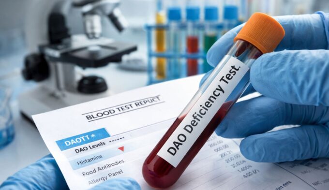
- DAO testing
- food-specific IgG panels
- stool or microbiome testing
- commercial “MCAS genetic panels”
- nonspecific inflammatory markers
- chromogranin A
- unvalidated heparin assays
These tests should be judged by the question they can actually answer. None has been validated as standalone diagnostic proof of MCAS. A random tryptase without a baseline comparison, for example, cannot establish whether an event-related rise occurred.
DAO testing is sometimes discussed in the evaluation of suspected histamine intolerance, but it does not diagnose systemic mast-cell activation or MCAS. Food-specific IgG panels and unvalidated heparin assays likewise do not provide standalone evidence of MCAS.
The key point is that none of these tests independently establishes mast-cell activation syndrome.
Why do MCAS diagnostic criteria differ online?
There are genuinely competing published frameworks for diagnosing MCAS, which explains a lot of the conflicting information you might encounter.
The commonly used ECNM-AIM allergy-immunology consensus approach, the framework this article anchors its practical guidance to, emphasizes severe recurrent systemic episodes, objective event-related biochemical evidence, and treatment response together. A separate, broader framework known as “consensus-2” was published in 2020 and updated again in 2026.
Consensus-2 uses wider diagnostic concepts than the stricter approach, and its proponents argue that more restrictive criteria risk missing genuine patients. Supporters of the stricter consensus criteria, in turn, emphasize diagnostic specificity and concern about potential overdiagnosis.
The two published frameworks remain distinct and should not be blended into a hybrid diagnostic system. For practical guidance, this article follows the ECNM-AIM criteria because they are used in the AAAAI work-group report and reflected in current practical clinical reviews. Consensus-2 is presented here as a separate, broader published framework.
What else may need to be ruled out?
Depending on how someone actually presents, a clinician may need to consider several other explanations before, or alongside, an MCAS evaluation. These can include IgE-mediated allergy and anaphylaxis, systemic mastocytosis or another clonal mast-cell disorder, chronic spontaneous or inducible urticaria, hereditary alpha-tryptasemia, medication reactions, POTS or other autonomic disorders, gastrointestinal disorders, and endocrine or neuroendocrine causes of flushing or rapid heart rate where clinically appropriate.
Not every patient needs testing for every item on this list; the actual symptom pattern guides which alternatives are worth investigating. MCAS also isn’t simply “a diagnosis of exclusion” in the sense of ruling everything else out first and defaulting to MCAS by elimination.
Appropriate alternative explanations need genuine consideration alongside a positive evaluation for the actual MCAS criteria, not instead of it. MCAS vs histamine intolerance covers one of the most common overlapping diagnostic questions in more depth.
How to prepare for an MCAS testing appointment
A little preparation makes the whole process considerably smoother. It helps to document the timing of your episodes in detail beforehand, since reproducibility and timing are central to how this gets evaluated.
Ask in advance how and where an acute tryptase sample can realistically be obtained during a future episode, since this often requires quick access to a lab or emergency setting. If urine testing is part of the plan, follow that specific laboratory’s collection instructions precisely, since protocols genuinely vary between labs.
Bring a complete medication list to your appointment, and don’t stop any prescribed medication unless your clinician specifically instructs you to. None of this preparation should ever come at the cost of delaying emergency treatment if you’re having a severe reaction; testing can wait, but a genuine emergency can’t.
When symptoms need urgent assessment
MCAS-like episodes can overlap in appearance with anaphylaxis, and certain symptoms should never wait for testing or a specific diagnosis. These include difficulty breathing, severe wheezing, swelling of the tongue or throat, fainting, severely low blood pressure, rapidly progressing symptoms across multiple body systems, blue or gray lips, and any severe reaction occurring for the first time.
These situations call for emergency assessment immediately. Never delay emergency treatment in order to obtain a tryptase sample first; getting the sample, if possible, comes after ensuring safety, not before.
Bottom line
MCAS diagnosis rests on a specific three-part combination: recurrent systemic symptoms, objective biochemical evidence of mast-cell activation, and meaningful improvement with mediator-targeted treatment. No single test, whether tryptase, a urinary mediator, or a genetic panel, substitutes for that full combination.
Serum tryptase interpreted against a person’s own baseline remains the most established biochemical evidence of systemic mast-cell activation, while urinary mediators can provide additional evidence with less standardized interpretation. A broader Consensus-2 framework also exists, but its criteria differ from the ECNM-AIM approach and should not be blended with it.
If your situation doesn’t fit neatly into this picture, that’s a reason to work through it carefully with a clinician experienced in mast-cell disorders, not a reason to assume any single test result tells the whole story.
Frequently asked questions
A few direct answers to the questions people bring to this topic most often.
Is there a blood test for MCAS?
Serum tryptase compared with a person’s own baseline is the most established blood-based biomarker for systemic mast-cell activation. Objective biochemical evidence is required under the commonly used consensus framework, but tryptase is not the only possible mediator evidence and does not diagnose MCAS by itself.
What is the 20% + 2 tryptase rule?
It’s a formula comparing an acute tryptase level to a person’s baseline, requiring the acute value to be at least baseline multiplied by 1.2, plus 2 ng/mL. Meeting this threshold documents significant event-related mast-cell activation.
When should tryptase be tested after a reaction?
Practical guidance suggests drawing the acute sample as soon as feasible, ideally within about one to four hours of symptom onset. A separate baseline sample should be collected at least 24 hours after complete recovery.
Can MCAS occur with normal tryptase?
Yes. An acute result can remain within the laboratory reference range and still meet the event-related formula if the baseline is low. If tryptase does not document activation, appropriately timed urinary mediators may sometimes provide additional objective evidence, but the commonly used consensus framework still requires biochemical evidence of mast-cell activation.
What urine tests are used for MCAS?
N-methylhistamine, leukotriene E4, and 2,3-dinor-11β-prostaglandin F2α are commonly studied urinary mediator tests used in mast-cell activation evaluation. They can support tryptase findings but are less standardized in their interpretation.
Does high baseline tryptase mean MCAS?
Not by itself. An elevated baseline can occur with systemic mastocytosis, hereditary alpha-tryptasemia, other blood conditions, and some unrelated causes, and it doesn’t independently diagnose MCAS.
Does a tryptase above 20 mean mastocytosis?
No. A baseline above 20 ng/mL is only one minor criterion considered when evaluating for systemic mastocytosis, and further evaluation depends on the full clinical picture, not that single number.
Can antihistamine response diagnose MCAS?
No. Antihistamines help many other allergic and dermatologic conditions too, so a response to them isn’t specific enough to confirm MCAS on its own; it’s only one part of the broader diagnostic framework.
Is there a genetic test for MCAS?
Not for MCAS itself. Genetic testing such as KIT D816V or assessment for hereditary alpha-tryptasemia addresses related but distinct questions, mainly around clonal mast-cell disease or tryptase genetics, rather than diagnosing MCAS directly.
How is MCAS different from systemic mastocytosis?
Systemic mastocytosis is a clonal mast-cell neoplasm diagnosed using separate tissue, molecular, and clinical criteria. MCAS describes recurrent systemic mast-cell activation that meets clinical, biochemical, and treatment-response criteria and may occur in primary/clonal, secondary, or idiopathic contexts. The two conditions can overlap, but they are not interchangeable.
References
- Afrin LB, Ackerley MB, Bluestein LS, et al. Diagnosis of mast cell activation syndrome: a global “consensus-2”. Diagnosis (Berl). 2020;8(2):137-152. doi:10.1515/dx-2020-0005
- Afrin LB, Blitshteyn S, Bluestein LS, et al. Progress in mast cell activation syndrome: the global consensus-2 diagnostic criteria at six years. Diagnosis (Berl). 2026. doi:10.1515/dx-2026-0016
- Gülen T, Akin C, Bonadonna P, et al. Selecting the Right Criteria and Proper Classification to Diagnose Mast Cell Activation Syndromes: A Critical Review. J Allergy Clin Immunol Pract. 2021;9(11):3918-3928. doi:10.1016/j.jaip.2021.06.011
- Lee E, Picard M. Diagnosis and management of mast cell activation syndrome (MCAS) in Canada: a practical approach. Allergy Asthma Clin Immunol. 2025;21:49. doi:10.1186/s13223-025-00998-9
- Valent P, Akin C, Arock M, et al. Definitions, criteria and global classification of mast cell disorders with special reference to mast cell activation syndromes: a consensus proposal. Int Arch Allergy Immunol. 2012;157(3):215-225. doi:10.1159/000328760
- Valent P, Bonadonna P, Hartmann K, et al. Why the 20% + 2 Tryptase Formula Is a Diagnostic Gold Standard for Severe Systemic Mast Cell Activation and Mast Cell Activation Syndrome. Int Arch Allergy Immunol. 2019;180(1):44-51. doi:10.1159/000501079
- Valent P, Akin C, Hartmann K, et al. Updated Diagnostic Criteria and Classification of Mast Cell Disorders: A Consensus Proposal. HemaSphere. 2021;5(11):e646. doi:10.1097/HS9.0000000000000646
- Voelker D, Pongdee T. Urine Mast Cell Mediators in the Evaluation and Diagnosis of Mast Cell Activation Syndrome. Curr Allergy Asthma Rep. 2024;24(2):33-38. doi:10.1007/s11882-024-01128-y
- Voelker D, Pongdee T. Biomarkers in the diagnosis of mast cell activation. Curr Opin Allergy Clin Immunol. 2025;25(1):27-33. doi:10.1097/ACI.0000000000001046
- Weiler CR, Austen KF, Akin C, et al. AAAAI Mast Cell Disorders Committee Work Group Report: Mast cell activation syndrome (MCAS) diagnosis and management. J Allergy Clin Immunol. 2019;144(4):883-896. doi:10.1016/j.jaci.2019.08.023
- Weiler CR. Mast Cell Activation Syndrome: Tools for Diagnosis and Differential Diagnosis. J Allergy Clin Immunol Pract. 2020;8(2):498-506. doi:10.1016/j.jaip.2019.08.022